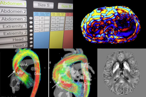
Techniques
CORE GIfMI supports a wide range of techniques, from structural scans to functional imaging and quantitative mapping, available for in-vivo and ex-vivo studies with healthy volunteers, patients, animals, and phantom experiments.
Looking for a specific technique or sequence? We can install additional sequences from the Siemens C2P platform. Contact our MR physicist for questions or custom setup.
Contact our MR Physicist
- Pim Pullens
- +32 9 332 89 75
- pim.pullens@ugent.be
Anatomical (structural) MRI Techniques
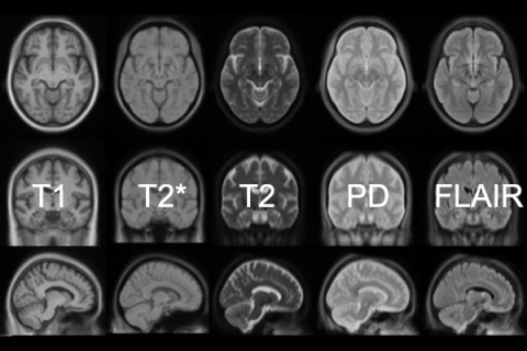
CORE GIfMI actively maintains a full library of optimized sequences, including:
- T1-weighted imaging
- T2-weighted imaging
- T2*-weighted imaging
- Fluid Attenuated Inversion Recovery (FLAIR)
- Proton density-weighted imaging
- 3D imaging: VIBE, SPACE, MP-RAGE, GRASP-VIBE, etc.
- and more
Functional MRI Techniques
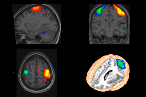
CORE GIfMI has extensive expertise in research using functional mapping techniques. Combined with the psychophyisological lab, these techniques enable wide-ranging opportunities for fMRI research:
- BOLD fMRI
- Simultaneous multi-slice (SMS) fMRI (Siemens and CMRR sequences)
- Task-based fMRI
- Resting-state fMRI
- and more
Quantitative MRI Techniques
The techniques are grouped into the following categories, according to the categorization described in Seiberlich, Nicole, Gulani, Vikas: Quantitative Magnetic Resonance Imaging. Academic Press; 2020. [Advances in Magnetic Resonance Technology and Applications, vol. 1].
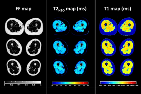
Relaxometry
- T1 mapping: Inversion recovery, Look-Locker, Variable Flip Angle, MP2RAGE
- T2 mapping: Multi-echo, Multi-spin echo, T2-prepared
- T2* mapping: Multi-echo, Multi-gradient echo
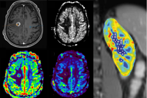
Perfusion and permeability
- Arterial Spin Labelling (ASL)
- Dynamic contrast-enhanced MRI (DCE) [not usable for healthy volunteers]
- Dynamic susceptibility contrast MRI (DSC) [not usable for healthy volunteers]
- Intravoxel incoherent motion (IVIM)
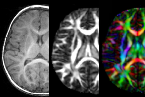
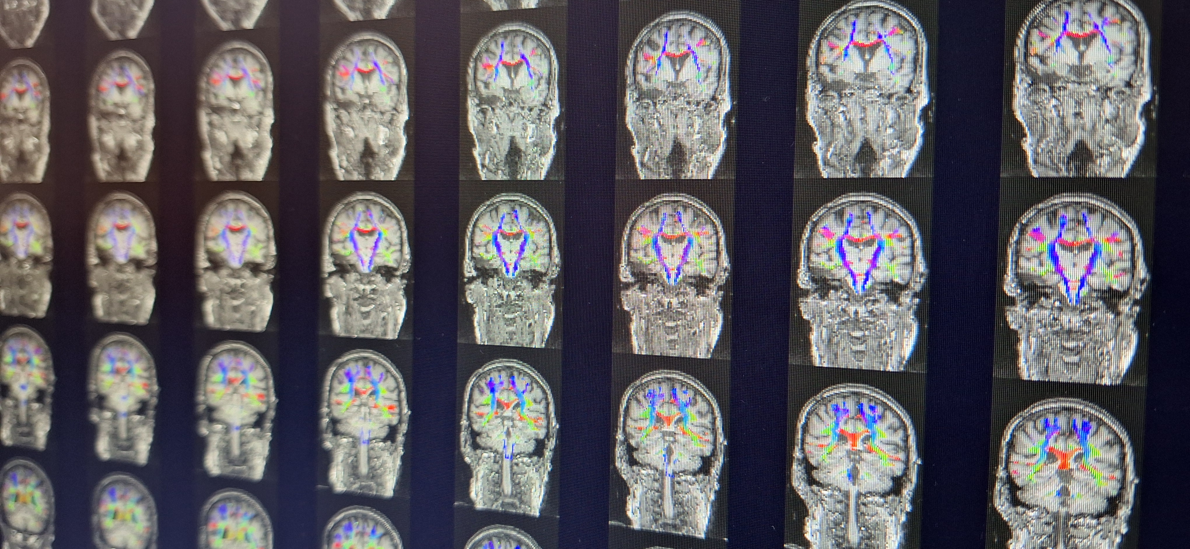
Diffusion
- ADC mapping
- Diffusion tensor imaging (DTI)
- High-angular resolution diffusion imaging (HARDI)
- Diffusion kurtosis imaging (DKI)
- Neurite orientation dispersion and density imaging (NODDI)
- Intravoxel incoherent motion (IVIM)
- Segmented diffusion-weighted imaging (RESOLVE)
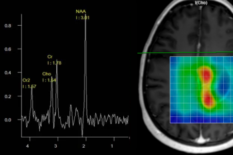
Spectroscopy
- Single-voxel
- Multi-voxel
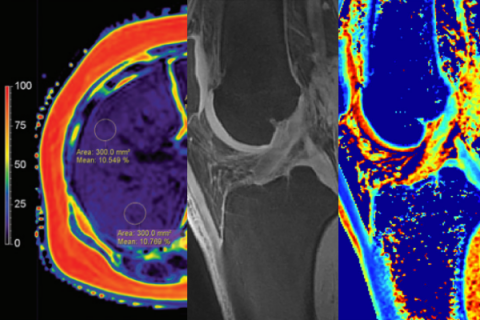
Fat and Iron quantification
- Proton Density Fat Fraction (PDFF)
- R2/T2 mapping
Quantification of Other MRI-Accessible Tissue Properties
- 2D Flow (phase contrast)
- 4D Flow
- Quantitative susceptibility mapping (QSM)
- Magnetic resonance elastography (MRE)
- Magnetization transfer imaging (MT)
- Chemical Exchange mapping (CEST)
- MR Thermometry
Deep Resolve

CORE GIfMI is equipped with the DeepResolve package from Siemens. DeepResolve is an advanced medical imaging technology that combines deep learning and artificial intelligence (AI) techniques to enhance the process of MRI image reconstruction, including several components:
- DeepResolve Gain: improves MRI signal-to-noise ratio by advanced denoising
- DeepResolve Sharp: enhances sharpness by a deep neural network
- DeepResolve Boost: accelerates MRI acquisition while maintaining high image quality by raw-data-to-image deep learning reconstruction technology
- DeepResolve Swift Brain: an ultra-fast brain protocol for T1, T2, T2*, FLAIR and DWI contrasts using deep learning based algorithms
Access to acquisition and reconstruction software
CORE GIfMI has on-scanner and offline access to multiple tools for sequence programming, image reconstruction and post-processing tools:
- Siemens C2P
The MRI pulse sequence sharing platform of Siemens Healthineers - Siemens IDEA
The sequence programming platform of Siemens Healthineers - Siemens ICE
The image reconstruction platform of Siemens Healthineers - Siemens OpenRecon
The inline image reconstruction platform of Siemens Healthineers - Pulseq
An open-source tool to design and share customizable MRI pulse sequences compatible with various scanners - gammaSTAR
A framework designed to create and deploy standardized MRI protocols across multiple scanner brands
Access to computation platforms and post-processing software
CORE GIfMI has access to multiple platforms for computing and data post-processing.
- HPC-UGent
The high performance computing infrastructure with lots of tools preinstalled and a huge computing power. - Neurodesk:
An environment running on the HPC for neuroimaging data analysis, see also our resources page. - Siemens syngo.sia Frontier
The Siemens Healthineers research platform
Quality Control
CORE GIfMI has specialized sequences and phantoms for MRI quality control, including: