Start a Study
Procedure from Start to Finish
Students and postdoctoral fellows are encouraged to submit proposals, with a faculty Principal Investigator (PI) as sponsor, participator and reviewer. All new studies are reviewed by the CORE GIfMI Board for their scientific value, innovation and feasibility.
I. Preparation of a new MRI study
1. Contact the CORE GIfMI Operational Team
An open conversation with the CORE GIfMI Operational Team (OPS) is the best way to begin a new project — especially if you’re new to MRI or have questions about:
- Getting started
- Scan parameters
- Study feasibility
- Special operational requirements
- Scan rates and budgeting
- Estimating scan time per participant (for informed consent)
We encourage you to schedule a kick-off meeting with the OPS Team to discuss your needs, address any concerns and to refine your experimental design. Please share the relevant literature well in advance so the Team can prepare and make the meeting as productive as possible.
2. Apply for Ethics Committee Approval
Human in vivo MRI scans may only be conducted after Ethics Committee approval. Submit academic and clinical research proposals via the HIRUZ (Health, Innovation and Research Institute) Research Portal.
- UZ Gent staff: Log in directly using your UZ institutional credentials.
- UGent staff, external collaborators, and students: Register for access.
Sections and wording to include in participant consent forms (text marked in bold-italic requires your specific input or adjustment):
MR Safety and Exclusion Criteria
Your safety is our top priority. Certain conditions or implants can make MRI scanning unsafe. Please review the following lists carefully before participating in an MRI study.
You CANNOT participate if you have any of the following:
- Cardiac pacemaker or defibrillator
- Retained wires from a removed electronic implant (e.g., pacemaker wires not attached to a device)
- Metal fragments in the eye or orbit
- Ferromagnetic aneurysm clip
- (Suspicion of) pregnancy
- Stainless steel intrauterine device (IUD)
You MAY NOT be able to participate if you have or have had:
- Artificial heart valve
- Ear or eye implant
- Brain aneurysm clip
- Implanted electronic device (e.g., drug infusion pump, electrical stimulator)
- Coil, catheter, or filter in any blood vessel
- Shrapnel, bullets, or other metal fragments
- Surgery, medical procedure, or tattoos (including permanent makeup) in the last six weeks
- Dental braces or major dental prostheses (depending on the scanned area)
- Severe claustrophobia (the scanner is a narrow tube)
Safety Review
All MRI research participants undergo thorough safety screening for implants or devices that might interact with the scanner’s magnetic field. If potential safety issues are identified, your case will be reviewed by the MRI Safety Responsible of the facility.
A decision about your participation will be made based on this assessment. In some cases, you may be asked to provide an operative report or implant details (such as device type and surgery date) to ensure safety.
Consent Form
Please ensure that a description similar to the one below, but accurate to your specific study procedure, is included in the consent form:
- Briefing - When you arrive at the MRI Facility, the researcher will clearly explain what the study involves and what will be expected of you. You’ll receive an overview of the MRI procedure to help you feel comfortable and well-informed. You will complete a detailed MRI Safety Screening Form, which will be reviewed with you by the MRI researcher to ensure your safety during the scanning session. There will be time for questions and discussion before the scan begins.
- Changing clothes – Your clothing - including underwear - must be completely free of metal parts and metal fibers (sometimes found in antimicrobial sportswear). You'll need to remove all metal items such as
- Glasses, hearing aids, or dentures
- Jewelry, watches, and hairpins
- Items from pockets (e.g., coins, paperclips, keys). These objects can be pulled toward the scanner magnet and may cause injury. It is recommended to wear long sleeves and long pants since the scanner room is relatively cool, unless the researcher provides other instructions in the context of the study.
- Scanning – (This section should reflect your specific study procedure)
- Example for a brain scan: The session begins with an anatomical brain scan (about 5 minutes), followed by several task-based scans, where you will respond to visual or auditory cues by pressing buttons. The total scan time is approximately 1 hour.
- Debriefing – After the scan, you'll be asked to complete a short post-MRI checklist about your experience. This helps the research team ensure participant comfort and improve future sessions.
Incidental Findings
This MRI examination is purely for research purposes. It is not medically indicated, is not a substitute for a clinical scan a doctor would order, and therefore has limited diagnostic value.
The design of the scan protocol may influence or even obscure abnormalities that a standard clinical MRI would detect. As a result:
- There is no routine review of scans for medical abnormalities by the researcher or a dedicated radiologist.
- No formal report will be generated.
If a researcher suspects an unexpected abnormality unrelated to the research question — which could range from minor anatomical variations to conditions requiring urgent medical attention — an expert radiologist will be consulted to:
- Confirm the finding
- Assess its clinical significance
- Recommend whether disclosure and referral to a physician are necessary
Any incidental findings will be communicated by a qualified medical professional, which could include:
- The researcher or Principal Investigator (if a medical doctor)
- Your general practitioner
- A relevant medical specialist
Please provide:
Your full name: ____________________________________
Phone number : __________________________
Family Doctor/Medical Clinic: ____________________________________________________________________
Your Rights and Data Protection
Your privacy and personal data are protected by law under:
- GDPR (EU) 2016/679, April 27, 2016
- Belgian law of July 30, 2018 on the protection of individuals regarding personal data
The researcher guarantees that
- Your privacy will be respected at all times
- You can request to access the data collected about you
- You can request correction of any errors in your data
- You can restrict the processing of your personal data
- You can request erasure of your data in certain circumstances
- You have the right to lodge a complaint
For more information on your rights and how to exercise them, visit the UGent website (https://www.ugent.be/nl/univgent/privacy/privacyverklaring.htm).
Data processing
By participation in this study, you consent to the processing or your data for research purposes. Only authorized personnel from the research team, under the responsibility of the Principal Investigator, will process your data. This may include internal staff without healthcare roles. The processing is lawful because it is:
- Performed in public interest (GDPR, Article 6, Paragraph 1e)
- Necessary for scientific research (GDPR, Article 9, Paragraph 2j)
Anonymity and Confidentiality
- All collected data will be pseudonymized: your data can only be linked to your personal identity via a code accessible to the investigator or their appointed delegate.
- Only pseudonymized data will be used for analysis, reports, publications, or presentations.
Data Storage
Your personal and health-related data will be retained for at least 10 years after study completion, for safety and follow-up reasons.
Data Sharing
- Pseudonymized data may be shared with future researchers for similar academic studies.
- Any re-use requires new Ethics Committee approval.
- If your data is transferred outside the European Economic Area (EEA), UGent ensures adequate protection through legal safeguards.
- Your explicit consent is required for international data transfers and is requested in the consent form below.
- If you do not want your data used for future research, you may contact the Data Protection Officer (DPO).
Contact details: privacy@ugent.be
Auditing
Representatives of the study promoter, auditors, the Medical Ethics Committee, and the competent authorities - all bound by professional secrecy - may access your study records. This access is strictly regulated by law and occurs under the responsibility of the investigator (or their authorized collaborator) to review study procedures and/or data. By signing this consent form, after receiving all preliminary explanations, you agree to this regulated access.
The Belgian supervisory Data Protection Authority, responsible for enforcing data protection legislation, can be contacted using the following contact details:
Data Protection Authority (DPA)
Rue de la Presse 35 – 1000 Brussels
Tel: +32 2 274 48 00
E-mail: contact@apd-gba.be
Website: www.dataprotectionauthority.be
II. While Ethics Approval is pending
3. Complete the MRI Study Proposal Form
The MRI Study Proposal Form helps the OPS Team to prepare for a smooth study launch (brief description, MR protocol, scan time needed, funding).
Send it to Stephanie.Bogaert@uzgent.be and Pieter.Vandemaele@ugent.be .
4. Gain Knowledge
While awaiting Ethics Committee approval, boost your MRI and post-processing knowledge. Setting up your post-processing pipeline in advance - before the large-scale acquisition of MRI data - can streamline your acquisition protocol or even eliminate unnecessary MRI sequences.
Recommended Resources:
- From Picture to Proton: A rigorous guide to fundamental MRI physics and technology.
- Doctoral Schools recordings: The GIfMI Ops team teaches Basic MRI Physics on a yearly basis.
- /documentationGIfMI Documentation Site: Collection of policies, forms and manuals to perform MRI research at CORE GIfMI.
- GIfMI Scanner User Manual: A must-read practical guide to help researchers get familiar with the MRI workflow and navigate the interface.
- GIfMI GitHub: Collection point for advanced questions and knowledge concerning MRI hardware and peripheral equipment.
- Siemens Healthineers Academy: Siemens offers a wide ranges of practical training videos,
- Some videos are freely accessible while others require creation of a login.
- Use Siemens Magnetom System Identifier 017-000166-73 for registration.
Siemens Healthineers Academy: relevant videos
System Overview
- Dual Monitor Set-up
- System Messages and Message History
- Scan UI Online self test (todo after having watched all other videos)
Routine Workflow
- System start-up and Home-screen
- Patient Registration
- Program Selection
- Scheduler
- Exam Workflow
- Job Aids (PDF with info on specific topics)
Post-processing and Reading
- MR View&GO Basic Online Training
- MR View&GO Advancend Online Training
- Configuration
- Reset Layout Function
- 3D Reference Point
- Distribution
- Patient Browser
- MR View&GO Online Self-Assessment
5. Join the MRI Community
It is highly recommended to join the CORE GIfMI communities.
- Our newsletter provides essential updates from the CORE GIfMI staff to all users. It is a one-way communication channel and is sent a few times per year.
- Join the Microsoft Teams space to connect, share ideas, ask questions, and collaborate.
- A quick way for users to share scan slots and updates is to join the WhatsApp group. The OPS Team is not included.
6. Attend the MRI Safety Training
All new users, including assisting Master’s students, must complete the free MRI Safety Training before participating in MRI research at CORE GIfMI.
Have you previously done an MRI project at our or another center? Contact the MRI Safety Responsible to discuss your training needs.
III. Study Proposal Approval
7. Provide Ethics Approval Information
Please complete the missing Ethics Committee Approval information in the MRI Study Proposal Form and submit a copy of the Subject Consent Form.
Required details:
- Ethics Committee Number
- Approval Date
- Expiration Date
- Upload Ethics Committee Approval Letter (pending)*
- Upload Informed Consent Form (pending)*
*Note - For now, documents can be sent by email to the Research Assistant.
8. Sign and Return the Rate Agreement (pending)
After study approval, the Principal Investigator must sign the Rate Agreement (pending). Your study can only start once CORE GIfMI has received the signed agreement. In return, the OPS Team will assign a GSB number for your study. This number is required for bookings and billing.
External users (non-UGent) are advised to contact Pim Pullens as soon as possible. A collaboration agreement (TechTransfer) must be set up, which takes time.
Rates
CORE GIfMI operates on a cost-recovery basis, with rates based on client type, staff, maintenance and resources. For current fees, cancellations, policy, and budget planning, see the Rates page.
Tip: use the Rate Calculator for your budget estimation. Contact the OPS Team for any further questions.
9. Request Access to the Booking System (CFMS)
The Core Facility Management Software (CFMS) is an online tool hosted by FD ICT at Ghent University to manage labs, resources, bookings, and billing. You can book MR scan time once you have
- Ethics Committee approval
- Approved MR Study Proposal
- Signed Rate Agreement
- GSB number
Main steps in using the CFMS:
- Every Principal Investigator (PI) conducting MRI experiments at CORE GIfMI must register in CFMS.
- Lab members handling data acquisition must register under their PI's lab and can book the facility with the PI’s approval.
- The PI must assign an account to the study to debit money from, see Adding Cost centers to a Lab in CFMS.
10. Request CORE GIfMI Facility Access
Access is only granted to MRI Safety trained individuals. You will need both a key and a badge to enter the facility.
Applying for a Facility Access Badge
This procedure applies to both UGent and external personnel:
- Contact the MRI Safety Responsible, who will coordinate with UZ Gent to request a cooperation with UZ Gent.
- Provide: work address, phone, email, preferred language, start date (annual renewal), and whether you have a Belgian eID or not.
- You will receive a registration link from the UZ Gent contractor portal; after registration your badge will be created.
- You will be notified by email when and where your badge is ready for pick-up. A deposit of 30 EUR is required (arranged mutually between UGent en UZ Gent).
Applying for a Facility Access Key
- Contact the MRI Safety Responsible who will coordinate with UZ Gent to request a key.
- Provide: work address, phone, email.
- Key production takes a few days.
- You will be notified by email when and where your key is ready for pickup. A deposit of 30 EUR is required (refunded upon return at project end).
11. Present your fMRI study
Note - this section does not apply to structural imaging studies.
Presenting your fMRI design early is optional but highly recommended. Early feedback from the fMRI community can help refine your design, improve data quality, and reduce issues during MRI acquisition and analysis.
Presentation format (15 - 20 minutes):
- Brief introduction: experimental hypothesis and research questions
- Detailed description of your experimental design
- Overview of expected results and how they will support your hypothesis
Registration: contact the fMRI support postdoc.
IV. To the Magnet
12. Set-up your MRI protocol
CORE GIfMI maintains a library of tested MRI sequences and protocols across various research domains.
Your scan protocol can be:
- Built from existing sequences with minor parameter adjustments, or
- Created from scratch for more complex studies
Need help designing your protocol? Book an appointment with the OPS Team.
13. Technical setup
CORE GIfMI provides a variety of peripheral equipment for MRI research — see the Equipment page for details.
- fMRI users: Test your paradigms thoroughly outside the scanner before pilot scans to minimize issues with triggering, stimulus-response acquisition, and log files.
- Other studies: Technical challenges may arise — the OPS Team can assist.
Book support: Contact the Support Engineer to schedule help with:
- Technical testing of peripheral equipment (triggers, button boxes, etc.)
- Any other technical questions
Note: If you plan to use non-standard peripheral equipment, especially in the MRI room, contact the Support Engineer beforehand.
14. Pilot Scans & Seed Funding
Pilot scans serve as a general rehearsal before actual data acquisition. They help to:
- Identify and resolve potential technical issues
- Asses scan time per subject
- Assess image quality
- Test in-scanner presentations
Note: Scanning a healthy volunteer (not part of the study) is allowed after Ethics Committee approval. For assistance, book an appointment with the OPS Team.
CORE GIfMI offers a seed funding program to promote the use of the GIFMI Core Facility and stimulate new MRI research. Successful applicants receive a limited number of hours free of charge.
Visit the seed funding program section to learn more.
15. Learn to operate the MRI scanner
Gain scanning experience under the guidance of experienced operators in your department or through on-site training with the OPS Team. Once comfortable with the equipment and participant interactions, you may scan independently.
Prepare well to accelerate your learning curve - scanner time is costly. The researcher is responsible for selecting a suitable a participant (non-claustrophobic and MRI-safe).
CORE GIfMI has developed key preparation materials. Please review the following documents before your first hands-on operator training:
16. Develop an SOP and checklist
Creating a practical Standard Operating Procedure (SOP) tailored to your project is key to capturing essential knowledge. Putting together a checklist ensures nothing's missed in any scan session.
17. Develop and test the post-processing pipeline
Don't wait until all data acquisition is complete - analyze data throughout your study as you go. Early development and testing of your pipeline helps to spot issues, optimize processing, and ensure data quality.
Key Data Management Tips:
- Data storage
- Define what to store: raw images, phsyio data, log files, etc.
- Plan storage for short-term (HDD, local servers, HPC) and long-term (cloud, repositories).
- During Data Acquisition
- Assess data quality per subject
- Run your processing pipeline per subject
- Review the output
- Perform interim group analysis if applicable
- Quality Assurance (QA)
- Set clear quality criteria
- Select appropriate QA tools like MRIQC (for fMRI) and visual inspections
- Combine automated and manual QA to catch acquisition artifacts and errors
- Data Analysis Resources
- Choose the right software for your analysis workflow (FSL, SPM, FreeSurfer, custom scripts)
- Ensure your have the necessary hardware (PC, a local server, HPC)
18. Start to Scan
You are now ready to scan the cohort.
Quick reminders:
- During acquisition, keep all scan parameters unchanged.
- Don't exceed approved subject count - get an ethics ammendment to add more.
- Use these MRI Safety Screening Forms in human imaging sessions.
- MR Safety Training for Students
All assisting students must complete the MRI Safety Training before accessing the facility, without exceptions, unless you get approval from the MRI Safety Responsible. - Booking cfms
- Please enter the fields according to the booking procedure described here to avoid manual corrections and implications on invoicing.
- Subject: enter GSB 99.999 – Doe John (space after GSB + dash flanked by spaces)
- Secial instruction: GSB 99.999 (space after GSB) - other comment should go on the second line
- Please enter the fields according to the booking procedure described here to avoid manual corrections and implications on invoicing.
- Data Storage
- MRI data is stored on the scanner for 9 days, then deleted by the GIfMI Operational Team.
- Export data to your own HDD.
- Incidental Findings
- Research scans aren't medical examins and won't be formally reviewed by a radiologist unless you suspect and report unexpected findings to the OPS Team. If needed, a radiologist will review the images and if clinically relevant, will refer the participant to a physician.
- Never share your own suspicions with participants - they may be incorrect and cause unnecessary worry.
- Equipment and Safety Reporting
- Report any equipment issues or safety incidents immediately to the MRI Safety Responsible.
V. After your MRI study
19. What after finishing a Study
- Notify the OPS Team when your last subject is scanned.
- Return your badge and/or key to the Badge Shop (Entrance 11) for a deposit refund. At the badge shop you will find a mailbox and envelopes to deposit your badge and key in. Write the relevant information on the envelope: name, address, IBAN bank account number, mobile phone number and email address. The personnel of the Badge Shop (09 332 94 09 - badgeshop@uzgent.be) will take care of the reimbursement.


- The OPS Team may request to document information to help future researchers benefit from your work.
20. Citation Guidelines for Scientific Output
Always acknowledge CORE GIfMI in publications and send us a copy of your work.
In scientific publications
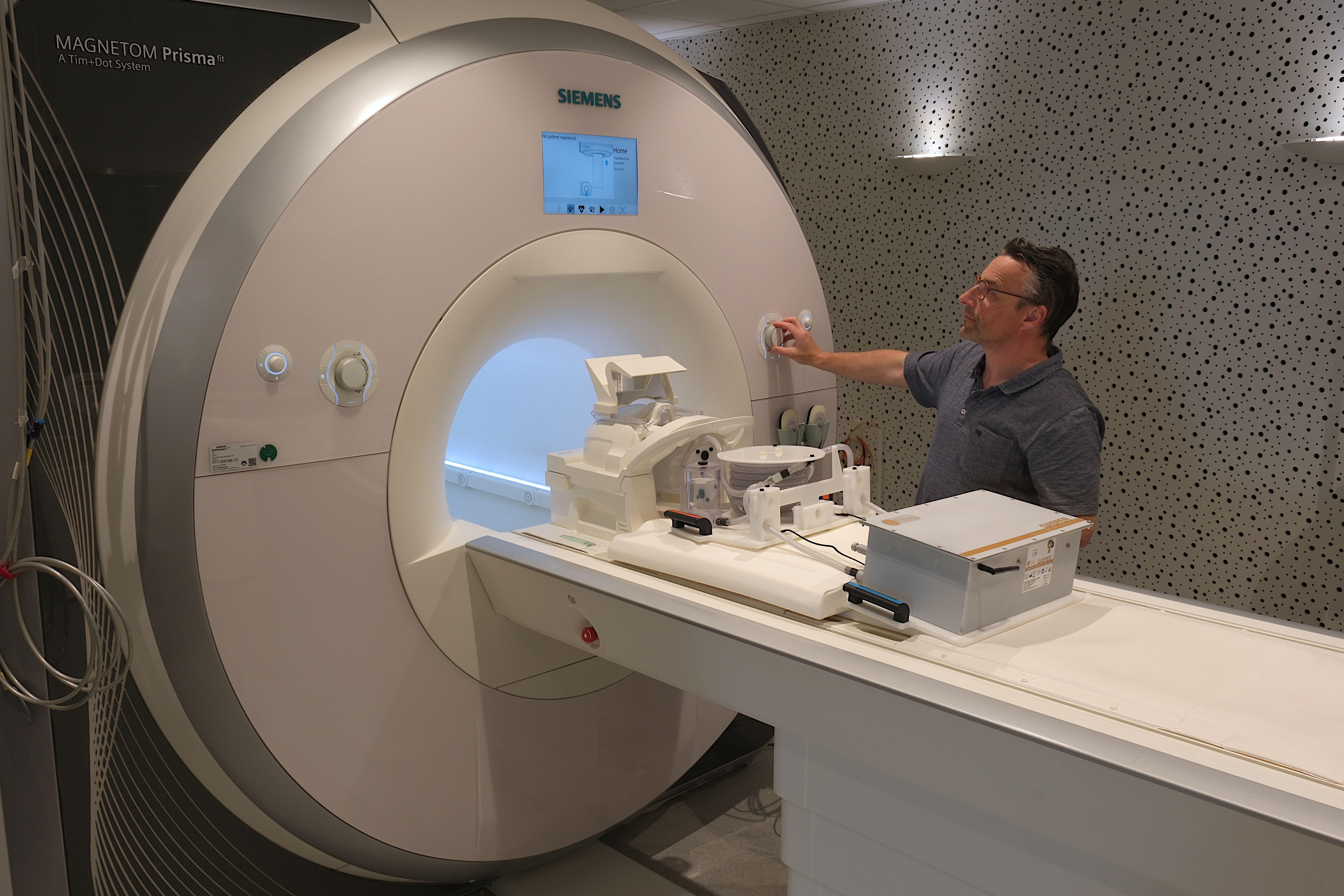
Materials and methods: MRI data was acquired at the Ghent Institute for Functional and Metabolic Imaging Core Facility (CORE GIfMI) at Ghent University (Belgium) using a 3T Siemens PrismaFit (Siemens Healthineers, Erlangen) MRI scanner.
Acknowledgements:
“The authors gratefully acknowledge [name(s) of the personnel] of the GIfMI Core Facility at Ghent University (Belgium) for the use and support on the 3T Siemens PrismaFit (Siemens Healthineers, Erlangen) MRI scanner."
With Core Facility staff as co-authors
Core Facility staff provide essential services to users, and their contribution may meet the criteria for co-authorship as defined by the International Committee of Medical Journal Editors (ICMJE). It is recommended to include the Core Facility as a separate affiliation in the author’s affiliation.
Example:
“First name Last name¹
GIfMI Core Facility, Ghent University, Ghent, Belgium”
On scientific posters and in presentations
“GIfMI Core Facility at Ghent University (Belgium)”