Techniques
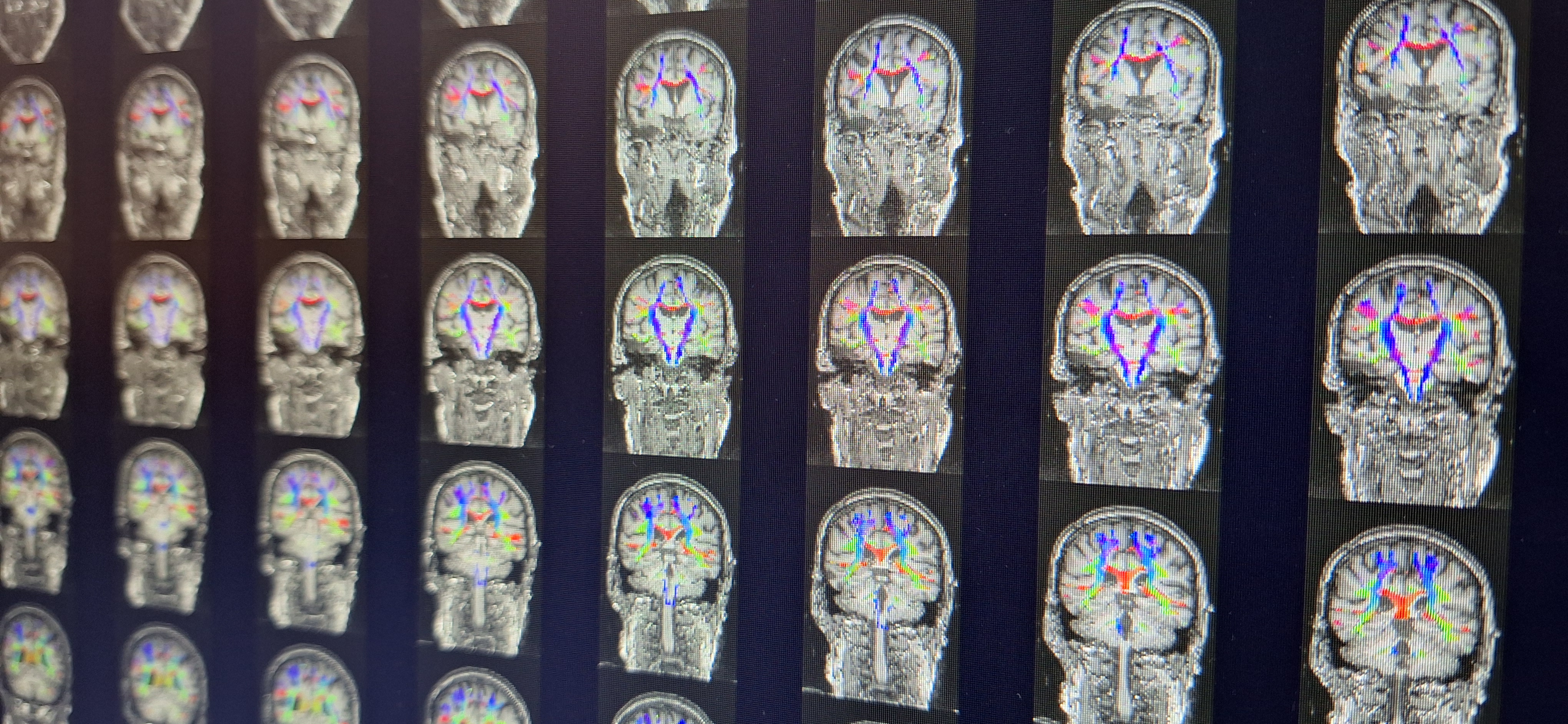
CORE GIfMI supports a wide range of techniques, from structural scans to functional imaging and quantitative mapping, available for in-vivo and ex-vivo studies with healthy volunteers, patients, animals, and phantom experiments.
Looking for a specific technique or sequence? We can install additional sequences from the Siemens C2P platform. Contact our MR physicist for questions or custom setup.
Contact our MR Physicist
- Pim Pullens
- +32 9 332 89 75
- pim.pullens@ugent.be
Anatomical (structural) MRI Techniques
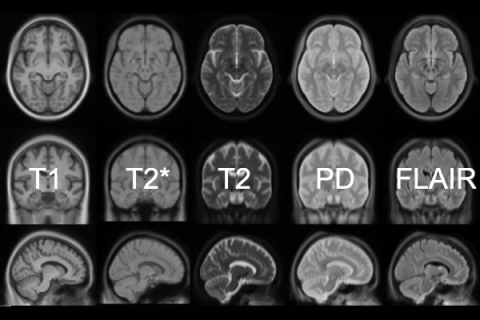
CORE GIfMI actively maintains a full library of optimized sequences, including:
- T1-weighted imaging
- T2-weighted imaging
- T2*-weighted imaging
- Fluid Attenuated Inversion Recovery (FLAIR)
- Proton density-weighted imaging
- 3D imaging: VIBE, SPACE, MP-RAGE, GRASP-VIBE, etc.
- and more
Functional MRI Techniques
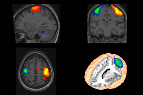
CORE GIfMI has extensive expertise in research using functional mapping techniques. Combined with the psychophyisological lab, these techniques enable wide-ranging opportunities for fMRI research:
- BOLD fMRI
- Simultaneous multi-slice (SMS) fMRI (Siemens and CMRR sequences)
- Task-based fMRI
- Resting-state fMRI
- and more
Quantitative MRI Techniques
The techniques are grouped into the following categories, according to the categorization described in Seiberlich, Nicole, Gulani, Vikas: Quantitative Magnetic Resonance Imaging. Academic Press; 2020. [Advances in Magnetic Resonance Technology and Applications, vol. 1][1].
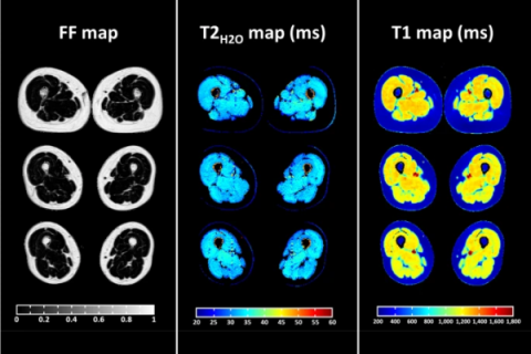
Relaxometry
- T1 mapping: Inversion recovery, Look-Locker, Variable Flip Angle, MP2RAGE
- T2 mapping: Multi-echo, Multi-spin echo, T2-prepared
- T2* mapping: Multi-echo, Multi-gradient echo
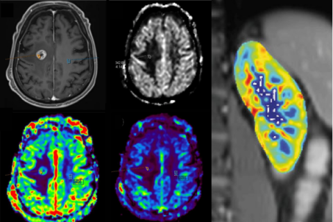
Perfusion and permeability
- Arterial Spin Labelling (ASL)
- Dynamic contrast-enhanced MRI (DCE) [not usable for healthy volunteers]
- Dynamic susceptibility contrast MRI (DSC) [not usable for healthy volunteers]
- Intravoxel incoherent motion (IVIM)
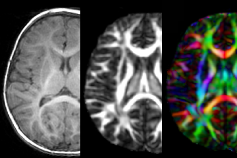
Diffusion
- ADC mapping
- Diffusion tensor imaging (DTI)
- High-angular resolution diffusion imaging (HARDI)
- Diffusion kurtosis imaging (DKI)
- Neurite orientation dispersion and density imaging (NODDI)
- Intravoxel incoherent motion (IVIM)
- Segmented diffusion-weighted imaging (RESOLVE)
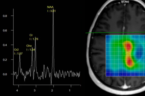
Spectroscopy
- Single-voxel
- Multi-voxel
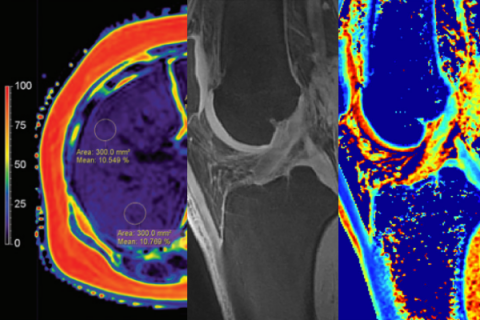
Fat and Iron quantification
- Proton Density Fat Fraction (PDFF)
- R2/T2 mapping
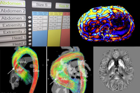
Quantification of Other MRI-Accessible Tissue Properties
- 2D Flow (phase contrast)
- 4D Flow
- Quantitative susceptibility mapping (QSM)
- Magnetic resonance elastography (MRE)
- Magnetization transfer imaging (MT)
- Chemical Exchange mapping (CEST)
- MR Thermometry
Access to acquisition and reconstruction software
CORE GIfMI has on-scanner and offline access to multiple tools for sequence programming, image reconstruction and post-processing tools:
- Siemens C2P
The MRI pulse sequence sharing platform of Siemens Healthineers - Siemens IDEA
The sequence programming platform of Siemens Healthineers - Siemens ICE
The image reconstruction platform of Siemens Healthineers - Siemens OpenRecon
The inline image reconstruction platform of Siemens Healthineers - Pulseq
An open-source tool to design and share customizable MRI pulse sequences compatible with various scanners - gammaSTAR
A framework designed to create and deploy standardized MRI protocols across multiple scanner brands
Access to computation platforms and post-processing software
CORE GIfMI has access to multiple platforms for computing and data post-processing.
- HPC-UGent
The high performance computing infrastructure with lots of tools preinstalled and a huge computing power. - Neurodesk:
An environment running on the HPC for neuroimaging data analysis, see also our resources page. - Siemens syngo.sia Frontier
The Siemens Healthineers research platform
Quality Control

CORE GIfMI has specialized sequences and phantoms for MRI quality control, including: